
Part VI — The Role of the Masquelet Technique in a Multistage Reconstruction Strategy
Drimadhariri.com
Introduction
Massive bone defects in the long bones represent one of the most challenging problems in reconstructive surgery. As the size of the defect increases, relying on a single technique to achieve all treatment objectives becomes increasingly difficult. In many cases, several goals must be addressed simultaneously: restoring the biological environment, recovering limb length, achieving mechanical stability, replacing the missing bone volume, controlling infection when present, and managing associated soft-tissue, vascular, or neurological injuries. [2,3,16,22–25]
Several techniques are available for managing these defects, including bone grafting, vascularized bone grafts, bone transport based on the principles of Ilizarov, and the Masquelet induced membrane technique. Each approach has its own advantages and limitations. [2,12–16,20–25]
When a bone defect exceeds 15 cm, a single technique may struggle to fulfil all the requirements of reconstruction. Reviews of the Masquelet technique have nevertheless documented its use in extremely large defects, including those exceeding 15 cm. [2]
This leads to the concept of hybrid reconstruction: combining more than one technique within a unified treatment strategy, allowing each method to address the component of the problem for which it is best suited.
In this article, I present two clinical cases involving exceptionally large bone defects: a 20 cm femoral defect and an 18.5 cm tibial defect. These cases provide an opportunity to discuss three key questions:
-
- Can a hybrid strategy be used to reconstruct bone defects exceeding 15 cm?
- What role can the Masquelet technique play as the biological foundation of a multistage reconstruction strategy?
- How do clinical observations align with cellular and biological studies suggesting that the induced membrane retains osteogenic properties beyond the conventional 6–8-week interval? [2,7,8]
Why Might a Hybrid Strategy Be Necessary?
When a bone defect is relatively small, reconstruction using bone grafting alone may be feasible.
However, as the size of the defect increases, the amount of graft required becomes substantial. Revascularization and the maintenance of graft viability also become major biological challenges. [2,3]
Bone transport, on the other hand, can regenerate new bone progressively and restore limb length. Nevertheless, it requires a prolonged treatment period and may be associated with complications, including pin-related problems, joint stiffness, axial deviation, and difficulties at the docking site. [12–16,22,23]
The Masquelet technique offers a different advantage: it creates a biological environment surrounded by an induced membrane that can support bone reconstruction. However, using this technique alone for extremely large defects may require substantial quantities of autologous bone graft. [1–3,5,7,8]
This raises an important surgical question:
Rather than selecting a single technique, why not use each technique to address the component of the problem that it is best suited to solve?
The Masquelet Technique as the Biological Foundation of Reconstruction
In a hybrid strategy, the objective is not necessarily to make the Masquelet technique responsible for replacing the entire bone defect.
Instead, its primary role may be to establish a favourable biological environment through the formation of an induced membrane. [1–8]
The first stage involves thorough debridement of non-viable bone and tissues, followed by placement of a cement spacer within the defect and appropriate stabilization of the limb. An induced membrane subsequently forms around the cement spacer. [1–3,5]
This membrane is not merely a mechanical envelope. It represents a biological environment containing blood vessels, cells, and factors involved in bone formation and revascularization. [5–11]
Studies have shown that the induced membrane releases important growth factors, including vascular endothelial growth factor (VEGF) and bone morphogenetic proteins (BMPs), and contributes to revascularization and osteogenesis. [5–8,10,11]
A hybrid reconstruction strategy may therefore follow this sequence:
Masquelet technique
↓
Formation of the induced membrane
↓
Bone transport / distraction osteogenesis
↓
Completion of the remaining defect using bone grafting or vascularized bone reconstruction, according to the clinical situation
In this approach, the techniques are complementary rather than competing alternatives.
Why Does the Problem Become More Challenging When the Defect Exceeds 15 cm?
When dealing with defects measuring 18.5 cm or 20 cm, the problem is no longer simply a matter of filling an empty space in the bone.
Such defects represent substantial losses that must be addressed from three perspectives:
- Biological reconstruction.
- Mechanical stability.
- Functional restoration.
In these situations, distributing the reconstructive tasks among several techniques may be more rational than expecting a single method to reconstruct the entire defect. [2,12–25]
This was the principle applied in the following two cases.
Case 1: A 20 cm Femoral Bone Defect Following Resection of a Malignant Bone Tumour
The first case involved a young, athletic, 23-year-old patient who presented with progressively increasing pain in the right thigh.
Clinical examination, imaging, and biopsy established the diagnosis of high-grade fibroblastic osteosarcoma.
Magnetic resonance imaging demonstrated a destructive intramedullary lesion. Computed tomography showed no distant metastases, and there was no direct involvement of the major femoral vessels.
(Figures 1 and 2)
Wide Resection and the First Stage
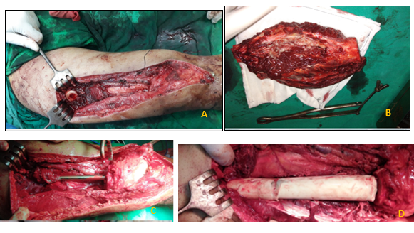
A wide tumour resection was performed, removing approximately 20 cm of the femoral shaft, together with the surrounding soft tissues, while preserving the neurovascular bundle.
The femur was stabilized using a retrograde interlocking intramedullary nail. PMMA bone cement was then placed within the defect to act as a spacer and stimulate the formation of the induced membrane, thereby completing the first stage of the Masquelet technique. [1–3,5]
(Figure 3)
Why Was the Second Stage Delayed?
This is an important aspect of the case.
After wound healing, the patient was referred to the oncology department and received six months of chemotherapy, according to the NCCN 2020 protocol.
Consequently, the second stage was delayed for approximately six months. This delay was not intended to investigate the longevity of the induced membrane. Rather, it was required to allow completion of the oncological treatment before proceeding with reconstruction.
Following chemotherapy, the patient was reassessed. Multiple biopsies were obtained from the resection margins and surrounding tissues to exclude tumour recurrence and infection. All results were negative.
Second Stage: Bone Transport
At this stage, distraction osteogenesis using bone transport was selected.
A modified, uniplanar AO external fixation system was used to permit long-distance bone transport while maintaining mechanical stability.
The original intramedullary nail was replaced with a longer nail to facilitate guidance of the transport process.
During removal of the cement spacer, the induced membrane was carefully preserved to maintain its biological properties. [2,7,8]
Bone transport began after a latency period of approximately two weeks, at a rate of 1 mm per day. [12–16]
The transport process continued for approximately 6.5 months, resulting in the formation of approximately 17.5 cm of new bone.
(Figures 4–7)
Because the external fixation system had reached the limits of its use and the patient did not wish to have the frame repositioned, transport was stopped after 17.5 cm. A residual defect of approximately 2.5 cm remained.
Third Stage: Completion of the Defect Using Bone Grafting
A third operation was performed to reconstruct the remaining 2.5 cm defect.
Autologous bone graft harvested from the iliac crest was used to fill the residual gap. After removal of the intramedullary nail, fixation was achieved using an intercondylar plate.
Additional bone and soft-tissue biopsies were obtained during the procedure to monitor for tumour recurrence and infection. All results were negative.
The overall reconstructive sequence was therefore:
Masquelet technique → Bone transport → Iliac crest bone grafting
(Figure 8)
Union and Functional Outcome
Complete union was achieved within 15 months.
Three months after the final bone-grafting procedure, the patient was able to discontinue crutches and bear full weight.
At four-year follow-up, there was no evidence of local tumour recurrence or distant metastases. The patient was walking independently, bearing full weight, and had returned to activities of daily living.
The residual limb-length discrepancy was less than 2 cm.
However, severe knee stiffness persisted. This was attributed to the prolonged treatment period, the presence of the external fixation frame around the knee, the extensive bone and soft-tissue resection, and interruption of physiotherapy during the bone-transport phase.
The functional outcome, assessed using the Musculoskeletal Tumor Society (MSTS) score, was 21/30, or approximately 70%.
(Figure 9)
Clinical Case Gallery — Case 1
The images documenting this case are available in the clinical case gallery:
Clinical case gallery: Hybrid reconstruction of a 20 cm femoral bone defect
📝 استئصال ورم عظمي عفلي خبيث من الفخذ لشاب 23 سنة وتم تعويض النقص 20 سم بطريقة ماسكليه(الغشاء المحرض) مع التزليق العظمي وتركيب سفود فخذ راجع واخيرا تطعيم عظمي حرقفي (الصور حسب التسلسل الزمني لعلاج الحالة )
What Did We Learn from This Case?
There was no need to replace the entire 20 cm defect using bone grafting alone.
Instead, the reconstructive task was divided among three complementary techniques:
- Masquelet technique: Establishment of the biological environment through formation of the induced membrane.
- Bone transport: Reconstruction of 17.5 cm and restoration of most of the lost femoral length.
- Bone grafting: Completion of the remaining 2.5 cm defect.
Bone transport achieved an external fixation index of approximately 11.1 days/cm and a consolidation index of approximately 26 days/cm. These values were favourable in comparison with published studies. [12–16]
The locally modified AO external fixation system was particularly important because it enabled long-distance bone transport in a resource-limited setting.
Case 2: An 18.5 cm Tibial Bone Defect Associated with Chronic Infection
The second case was fundamentally different.
The patient was a 23-year-old man who had sustained a high-energy open tibial fracture classified as Gustilo–Anderson type IIIC, accompanied by severe soft-tissue damage and neurovascular injury.
Initial management included vascular reconstruction using a reversed saphenous vein bypass, followed by multiple surgical procedures involving debridement, fixation, and soft-tissue coverage.
Over approximately two years, the patient developed chronic osteomyelitis, bone necrosis, and extensive soft-tissue loss, with exposure of the plate and screws.
He also had incomplete injuries involving both the tibial and common peroneal nerves.
Following several previous attempts to treat the osteomyelitis, the segmental tibial defect measured 18.5 cm. The limb was severely unstable, and the patient was strongly considering below-knee amputation.
(Figures 1 and 2: The initial condition of the limb and its appearance when the patient presented for staged bone reconstruction.)
First Stage: The Masquelet Technique
Radical debridement of all necrotic and infected bone and soft tissues was performed.
Antibiotic-loaded PMMA cement was placed within the defect to stimulate induced-membrane formation. The limb was stabilized using a two-level external fixation system. [1–3,19]
(Figure 3)
As in the first case, proceeding directly to the second stage was not possible.
The interval was necessary to ensure adequate infection control and resolution of the active infectious process before continuing reconstruction. The period between the first and second stages was approximately six months.
Once again, this delay was not a deliberate experiment to investigate the age of the membrane. It was a clinical decision dictated by the patient’s condition.
Second Stage: Bone Transport
After maturation of the induced membrane, the cement spacer was removed and a modified AO bone-transport device was applied.
A corticotomy was performed to initiate distraction osteogenesis and bone transport. [12–16]
(Figures 4 and 5)
Bone transport continued for 162 days, achieving a length gain of 6.5 cm, at an average rate of approximately 0.4 mm per day.
The slower transport rate was related to severe soft-tissue contracture in the leg and the presence of a vascular bypass.
A modified bilateral tubular external fixation system was used instead of a conventional circular Ilizarov frame, as it was easier for the patient to tolerate.
Third Stage: Fibular Tibialization
After 6.5 cm of bone transport, further advancement became impossible because the surrounding soft tissues could no longer accommodate continued transport.
The procedure was therefore stopped at this point, leaving a residual tibial defect of approximately 12 cm.
Fibular tibialization was subsequently performed.
The ipsilateral fibula was transferred into the defect while preserving its blood supply, muscular attachments, and surrounding soft-tissue connections. This provided living bone with both biological and mechanical potential. [17,20,21]
The proximal end of the fibula was divided and transferred to the proximal tibial segment, while the distal end remained connected to the ankle joint. The transferred fibular segment was then fixed within the defect.
(Figures 6 and 7)
A limited area of superficial skin necrosis developed postoperatively. It was managed conservatively with local wound care and healed within two weeks without further surgery.
The final reconstructive sequence was:
Masquelet technique → Bone transport → Vascularized fibular reconstruction
Results
After 24 months, complete tibial reconstruction had been achieved, restoring the structural continuity of the limb.
Radiographs demonstrated formation of the regenerate bone followed by consolidation. There was also marked hypertrophy of the transferred fibula, consistent with Wolff’s law.
(Figure 8)
No recurrence of infection occurred during follow-up.
The patient regained independent walking with progressive weight-bearing and recovered the full range of knee motion. However, partial deficits in ankle and toe movements persisted as a consequence of the original nerve injuries.
(Figure 9)
A residual limb-length discrepancy of approximately 4 cm remained. This was compensated for using a shoe lift, with the possibility of further limb lengthening in the future if clinically indicated.
Considering the severity of the original injury and the magnitude of the bone defect, the functional outcome was satisfactory.
Clinical Case Gallery — Case 2
The clinical images are available in the second case gallery:
Clinical case gallery: Hybrid reconstruction of an 18.5 cm tibial bone defect
📝 صور الاشعة والحالة المرضية من بداية الاصابة حتى الشفاء الكامل وتعويض الضياع العظمي (الصور مرقمة حسب التسلسل الزمني)
What Do the Two Cases Have in Common?
Although the two cases differed substantially in their underlying pathology, both followed a similar reconstructive principle.
| Feature | Femoral defect | Tibial defect |
| Defect size | 20 cm | 18.5 cm |
| Cause | Resection of a malignant bone tumour | Severe trauma with chronic infection |
| First stage | Masquelet technique | Masquelet technique |
| Reason for delay | Completion of chemotherapy | Infection control |
| Interval between stages | Approximately 6 months | Approximately 6 months |
| Main reconstructive procedure | Bone transport | Bone transport |
| Completion of the residual defect | Iliac crest bone graft | Fibular tibialization |
| Outcome | Union and limb salvage | Reconstruction and limb salvage |
The shared feature was not simply the successful reconstruction of both defects.
Rather, it was the use of the Masquelet technique as the biological foundation, followed by bone transport and an additional reconstructive procedure selected according to the requirements of each case.
This approach is consistent with the literature describing the Masquelet technique as one of several strategies for reconstructing segmental bone defects. Distraction osteogenesis, bone transport, and vascularized bone grafting remain important alternatives or complementary procedures, depending on the nature of the defect and its biological and mechanical conditions. [2,12–25]
What Do Cellular Studies Tell Us About the Induced Membrane After Eight Weeks?
This question is important for interpreting the outcomes of the two cases.
Traditionally, the second stage of the Masquelet technique has generally been performed within 4–8 weeks, based on the concept that the induced membrane exhibits substantial biological activity during this period. [2,7]
However, subsequent cellular and histological studies have suggested that this timing window may be less rigid than previously assumed. [7,8]
The Study by Gessmann and Colleagues
In a study published in the European Journal of Trauma and Emergency Surgery, Gessmann and colleagues investigated induced membranes obtained from 60 patients, with membrane maturation periods ranging from one to 16 weeks. [7]
The investigators evaluated the membranes using:
- Histological examination.
- Co-culture with mesenchymal stem cells (MSCs).
- ELISA analysis of membrane extracts.
- Protein microarray analysis.
The study reported several findings:
- Vascularization increased in membranes older than four weeks.
- Fibrosis increased after approximately seven weeks.
- MSCs could be grown from all the membranes studied.
- All membranes promoted the proliferation of cultured MSCs.
- Osteocalcin and osteopontin were detected across the studied time points, without statistically significant differences. [7]
The principal finding relevant to reconstruction was that membranes older than eight weeks retained regenerative properties comparable to those of younger membranes. [7]
Therefore, structural changes associated with membrane maturation, including increased vascularization and fibrosis, do not necessarily indicate that the membrane has lost its biological capacity to support reconstruction. [7]
What Happens After Several Months?
The study by Gindraux and colleagues is particularly relevant to the two clinical cases presented here.
The investigators studied 34 patients and obtained membrane samples from seven patients between 4.2 and 14.7 months after the first stage.
The mean interval before the second stage was 5.8 months, with the longest interval reaching 14.7 months.
Analysis of the older membrane samples demonstrated the presence of:
Mesenchymal stromal cells with in-vitro osteogenic potential.
In other words, stromal cells capable of osteogenic activity in vitro remained present in the induced membrane. [8]
The study also indicated that the membrane represents a multipotent tissue retaining properties associated with osteogenic and chondrogenic differentiation. [8]
The authors concluded that the induced membrane can retain osteogenic properties over time and that a longer interval between the two stages was not associated with delayed bone healing. [8]
How Do These Findings Relate to Our Two Cases?
In the first case, the second stage was delayed for approximately six months because the patient needed to complete chemotherapy for the malignant tumour.
In the second case, the interval was also approximately six months, but the delay was required to ensure adequate control of the infection.
Thus, neither delay was deliberately introduced to test how long the induced membrane could remain biologically active.
Nevertheless, when the patients were able to proceed to the next stage:
- The induced membrane could be preserved.
- The membrane could be incorporated into the reconstructive strategy.
- Successful reconstruction was achieved in both cases.
These clinical observations are consistent with the published cellular and histological evidence. [7,8]
However, it is important not to claim that these two cases prove that the membrane retains exactly the same biological activity for six months.
A more accurate interpretation is:
Cellular and histological studies suggest that the induced membrane can retain biological and osteogenic properties beyond the conventional interval of 6–8 weeks. The two cases presented here provide clinical examples in which the membrane was used successfully after the second stage had been delayed for approximately six months because of treatment-related requirements. [7,8]
From Choosing a Technique to Designing a Strategy
These cases illustrate a shift in surgical thinking.
Instead of asking:
“Which technique should I use?”
The more useful question may be:
“Which combination of techniques best addresses this patient’s problem?”
Each technique can have a distinct role.
Masquelet Technique
Establishing the biological environment through formation of the induced membrane.
Bone Transport
Restoring limb length and reconstructing a substantial proportion of the defect.
Bone Grafting
Completing the residual bone defect when appropriate.
Vascularized Fibular Reconstruction
Providing living, vascularized bone when the biological and reconstructive requirements justify its use. [17,20,21]
In this way, different techniques become complementary tools within a single reconstructive strategy rather than mutually exclusive alternatives.
Important Technical Considerations
In the femoral case, bone transport was performed at 1 mm per day. The external fixation index was approximately 11.1 days/cm, and the consolidation index was approximately 26 days/cm. These values were favourable in comparison with published ranges. [12–16]
In the tibial case, a slower transport rate of approximately 0.4 mm per day was selected because of the previous infection and the compromised soft tissues.
The external fixation index was approximately 15 days/cm, and complete consolidation took approximately 24 months.
These differences emphasize that the bone-transport strategy itself can be adjusted according to the biological and mechanical conditions of the limb. [12–16,22,23]
Clinical Pearls
- Bone defects exceeding 15 cm may require a multi-technique reconstructive strategy rather than reliance on a single procedure. [2,12–25]
- The Masquelet technique can provide the biological foundation for reconstruction, while other techniques restore length and replace the remaining bone defect. [2,12–25]
- Exceeding the conventional 6–8-week interval does not necessarily mean that the induced membrane has lost its biological properties. [7,8]
- Cellular studies have demonstrated the persistence of MSCs and osteogenesis-related factors in membranes beyond eight weeks. Another study identified cells with osteogenic potential in membranes several months after the first stage. [7,8]
- Delaying the second stage may be necessary for clinical reasons, including completion of chemotherapy or confirmation of infection control.
- In both cases presented here, the interval between the two stages was approximately six months, yet successful reconstruction was achieved.
- Hybrid reconstruction is not a fixed protocol. The strategy must be tailored to the nature of the defect and the individual patient’s condition.
Conclusion
These two cases illustrate that reconstruction of massive bone defects may require a shift from the concept of a single technique to that of a hybrid reconstructive strategy.
In both cases, the Masquelet technique provided the initial biological foundation through formation of the induced membrane. Bone transport was then used to reconstruct a substantial portion of the defect and restore limb length. A different procedure was selected to complete the reconstruction in each case: bone grafting for the femoral defect and vascularized fibular transfer for the tibial defect.
The defects measured 20 cm in the femur and 18.5 cm in the tibia, yet successful reconstruction and limb salvage were achieved.
These cases suggest that dividing a massive defect into sequential reconstructive stages and assigning complementary roles to different techniques may provide a practical solution when a single method is insufficient. [2,12–25]
Furthermore, the second stage was performed after an interval of approximately six months in both cases, for different clinical reasons. These observations are consistent with cellular and histological evidence suggesting that the induced membrane may retain biological and osteogenic properties beyond the conventional 6–8-week interval. [7,8]
Therefore, the interval of 6–8 weeks should not necessarily be regarded as an absolute deadline. The timing of the second stage should be determined clinically, taking into account the patient’s condition, infection control, soft-tissue status, oncological treatment, and reconstructive plan. [7,8]
In massive bone defects, the best solution may not be a single technique, but a carefully designed strategy in which each technique performs the task for which it is best suited.
References
- Masquelet AC, Fitoussi F, Bégue T, Muller GP. Reconstruction of the long bones by the induced membrane and spongy autograft. Ann Chir Plast Esthet. 2000;45(3):346–353.
- Masquelet AC. Masquelet’s induced membrane technique: Review of current concepts and future directions. J Orthop Res. 2021;39(4):707–718.
DOI: 10.1002/jor.24978 - Pelissier P, et al. The induced membrane technique for the treatment of massive bone defects: a review. J Orthop Sci. 2013;18(5):690–698.
- Masquelet AC, Begue T. The Masquelet technique: A new method for bone reconstruction. Orthop Traumatol Surg Res. 2010;1(1):10–18.
- Aho OM, Lehenkari P, Ristiniemi J, Lehtonen S, Risteli J, Leskelä HV. The mechanism of action of induced membranes in bone repair. J Bone Joint Surg Am. 2013;95(7):597–604.
DOI: 10.2106/JBJS.L.00310 - Tang Q, Tong M, Zheng G, et al. Masquelet’s induced membrane promotes the osteogenic differentiation of bone marrow mesenchymal stem cells by activating the Smad and MAPK pathways. Am J Transl Res. 2018;10(4):1211–1219.
- Gessmann J, Rosteius T, Baecker H, Sivalingam K, Peter E, Schildhauer TA, Köller M. Is the bioactivity of induced membranes time dependent? Eur J Trauma Emerg Surg. 2022;48(4):3051–3061.
DOI: 10.1007/s00068-021-01844-4 - Gindraux F, Loisel F, Bourgeois M, et al. Induced membrane maintains its osteogenic properties even when the second stage of Masquelet’s technique is performed later. Eur J Trauma Emerg Surg. 2020;46(2):301–312.
DOI: 10.1007/s00068-019-01184-4 - Gindraux F, et al. Correction to: Induced membrane maintains its osteogenic properties even when the second stage of Masquelet’s technique is performed later. Eur J Trauma Emerg Surg. 2020;46(2):313–315.
DOI: 10.1007/s00068-019-01242-x - Henrich D, Seebach C, Nau C, et al. Establishment and characterization of the Masquelet induced membrane technique in a rat femur critical-sized defect model. J Tissue Eng Regen Med. 2016;10–E396.
DOI: 10.1002/term.1826 - Gouron R, Petit L, Boudot C, et al. Osteoclasts and their precursors are present in the induced-membrane during bone reconstruction using the Masquelet technique. J Tissue Eng Regen Med. 2014/2017;11(2):382–389.
DOI: 10.1002/term.1921 - Ilizarov GA. The tension-stress effect on the genesis and growth of tissues. Clin Orthop Relat Res. 1989;(238):249–281.
- Paley D. Problems, obstacles, and complications of limb lengthening by the Ilizarov technique. Clin Orthop Relat Res. 1990;(250):81–104.
- Kocaoglu M, Eralp L, Rashid HU, et al. Reconstruction of segmental bone defects due to chronic osteomyelitis with use of an external fixator and an intramedullary nail. J Bone Joint Surg Am. 2006;88(10):2137–2145.
DOI: 10.2106/JBJS.E.01152 - Li Z, Zhang X, Duan L, et al. Distraction osteogenesis technique using an intramedullary nail and a monolateral external fixator in the reconstruction of massive postosteomyelitis skeletal defects of the femur. Can J Surg. 2009;52(2):103–111.
- Mainardi L, et al. Distraction osteogenesis for the reconstruction of large bone defects: A systematic review and meta-analysis. Injury. 2022;53(6):2038–2046.
- Yin P, Zhang L, Li T, et al. Ipsilateral fibula transport for the treatment of massive tibial bone defects. Injury. 2015;46(11):2273–2277.
DOI: 10.1016/j.injury.2015.08.028 - Grün W, Hansen EJ, Andreassen GS, Clarke-Jenssen J, Madsen JE. Functional outcomes and health-related quality of life after reconstruction of segmental bone loss in femur and tibia using the induced membrane technique. Arch Orthop Trauma Surg. 2023;143(8):4587–4596.
DOI: 10.1007/s00402-022-04714-9 - Mathieu L, Mourtialon R, Durand M, et al. Masquelet technique in military practice: specificities and future directions for combat-related bone defect reconstruction. Mil Med Res. 2022;9(1):48.
DOI: 10.1186/s40779-022-00411-1 - Houben RH, Rots M, van den Heuvel SCM, Winters HAH. Combined massive allograft and intramedullary vascularized fibula as the primary reconstruction method for segmental bone loss in the lower extremity: a systematic review and meta-analysis. JBJS Rev. 2019;7(8).
DOI: 10.2106/JBJS.RVW.18.00166 - Petersen MM, Hovgaard D, Elberg JJ, et al. Vascularized fibula grafts for reconstruction of bone defects after resection of bone sarcomas. Scientifica. 2010;2010:524721.
DOI: 10.1155/2010/524721 - Quinnan SM. Segmental bone loss reconstruction using ring fixation. J Orthop Trauma. 2017;31(Suppl 5)–S46.
DOI: 10.1097/BOT.0000000000000985 - Plotnikovs K, Kamenska J, Movcans J, et al. Artificial deformity creation as a method for limb salvage for patients with massive tibial and soft tissue defects: a report of 26 cases. Strategies Trauma Limb Reconstr. 2023;18(3):133–139.
DOI: 10.5005/jp-journals-10080-1599 - Adamczyk A, Meulenkamp B, Wilken G, Papp S. Managing bone loss in open fractures. OTA Int. 2020;3(1).
DOI: 10.1097/OI9.0000000000000059 - Hayashi K, Futamura K, Ogawa T, et al. Management of bone loss in acute severe open tibial fractures: a retrospective study of twenty-nine cases—a treatment strategy with bone length preservation. Int Orthop. 2023;47(6):1565–1573.
DOI: 10.1007/s00264-023-05760-7
Related Articles in This Series
- Part I: The Masquelet technique for the management of large bone defects
- Part II: The induced membrane — histological structure and biological properties
- Part III: Indications for the Masquelet technique and patient selection
- Part IV: The Masquelet technique for the management of large bone defects
- Part V: The second stage of the Masquelet technique — cement removal, bone grafting, and reconstruction