
The Masquelet Technique for the Treatment of Massive Bone Defects – Part IV
Part IV: The First Stage of the Masquelet Technique – Surgical Principles and Practical Steps
By Dr. Imad Al Hariri
Consultant Orthopaedic Surgeon
Based on a lecture on the importance of the Masquelet technique in the reconstruction of massive bone defects, delivered at the Annual Orthopaedic Surgery Conference, 2018, Omayyad Hotel, Damascus.
Article type: Surgical Technique – Narrative Review
Introduction
The first stage of the Masquelet technique represents the foundation upon which all subsequent stages of treatment are built. The success of bone reconstruction during the second stage depends directly on the quality of the first-stage procedure.
Some surgeons believe that the purpose of this stage is limited to filling the bone defect with cement. However, this concept is inaccurate. In reality, this stage aims to achieve several surgical and biological objectives simultaneously, including the elimination of non-viable tissues, control of infection when present, restoration of mechanical stability of the limb, and preparation of a suitable environment for induced membrane formation.
Therefore, any error during this stage may lead to failure of reconstruction, regardless of how well the second stage is performed.
Contents
-
- Introduction
- Objectives of the First Stage
- Preoperative Planning
- Step One: Radical Surgical Debridement
- How Do We Determine the Limits of Debridement?
- Step Two: Preparation of the Bone Ends
- Step Three: Achieving Stable Bone Fixation
- What Fixation Methods Are Available?
- Step Four: Preparation and Application of Bone Cement
- When Should the Cement Be Applied?
- What Should the Shape of the Cement Be?
- Step Five: Adequate Soft-Tissue Coverage
- What Happens After Completion of the First Stage?
- Common Errors During the First Stage
- Conclusion
- References
Objectives of the First Stage
The objectives of the first stage can be summarised in five main goals:
-
- Removal of all non-viable tissues.
- Eradication of infection when present.
- Achievement of stable bone fixation.
- Preservation of the bone defect space.
- Stimulation of induced membrane formation.
The surgeon should not proceed to the next stage until these objectives have been fully achieved.
Preoperative Planning
Before starting the operation, the case should be carefully evaluated through:
-
- Assessment of radiographic images.
- Determination of the actual length of the bone defect.
- Evaluation of soft-tissue integrity.
- Assessment of the limb’s blood supply.
- Investigation for signs of infection.
- Selection of the appropriate fixation method.
The surgeon should also anticipate the possible need for muscle or skin coverage when there is extensive soft-tissue loss.
Step One: Radical Surgical Debridement
Surgical debridement is the most important step in the first stage.
Debridement must be extensive and radical, ensuring removal of all tissues that do not have adequate viability.
This includes:
-
- Fibrous tissue.
- Scar tissue.
- Necrotic bone.
- Foreign-body remnants.
- Infected tissues.
- All interposed tissues between the two ends of the bone defect.
How Do We Determine the Limits of Debridement?
Bone resection should continue until viable bone with good blood supply is reached.
The surgeon can use several intraoperative findings as indicators:
-
- The appearance of bleeding from the medullary canal and the cut bone surface, known as the Paprika Sign.
- Disappearance of grey, friable, or non-bleeding tissues.
- Achievement of firm bone on examination.
Leaving portions of non-viable bone may lead to persistent infection or subsequent failure of bone union.
Step Two: Preparation of the Bone Ends
After completion of debridement, both bone ends should be carefully cleaned and prepared.
The objectives are to:
-
- Remove adherent fibrous tissue.
- Remove non-viable bone layers.
- Expose viable bone.
- Provide a suitable surface for future bone healing.
Step Three: Achieving Stable Bone Fixation
After completing debridement, the bone must be stabilised securely.
Mechanical stability is one of the most important requirements for the success of the technique, because both the induced membrane and the bone graft require a stable environment to allow reconstruction to take place.
Fixation must be robust and should remain in place until final bone union has been achieved.
What Fixation Methods Are Available?
One of the advantages of the Masquelet technique is that it does not require a specific fixation method. Instead, the most appropriate method can be selected according to the nature of the injury.
Available fixation methods include:
-
- External fixation.
- Plate fixation.
- Intramedullary nailing.
- Hybrid fixation.
The choice depends on:
-
- The location of the bone defect.
- The presence of infection.
- The condition of the soft tissues.
- The patient’s age.
- The type of injury.
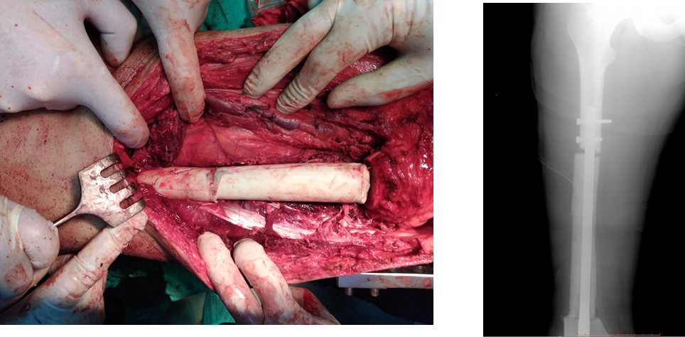
Figure 1. Filling the bone defect with cement after debridement of the bone ends and stabilisation of the bone using an intramedullary-supported rod.
Step Four: Preparation and Application of Bone Cement
After stabilising the bone, polymethylmethacrylate bone cement (PMMA) is prepared.
Its functions during this stage are to:
-
- Maintain the bone defect space.
- Prevent invasion of the area by fibrous tissues.
- Stimulate the formation of the biological membrane.
- Contribute to local infection control when antibiotic-loaded cement is used in appropriate cases.
When Should the Cement Be Applied?
Your lecture emphasised an important practical point: the cement should be applied immediately before it has completely hardened, so that it can be shaped easily to match the dimensions of the bone defect and fill the entire space.
The cement should fill the full length and width of the defect without leaving any gaps.
What Should the Shape of the Cement Be?
The role of the cement is not limited to filling the defect.
It should:
-
- Fill the entire bone defect.
- Maintain limb length.
- Prevent the entry of soft tissues.
- Allow easy removal during the second stage.
- Allow formation of a membrane surrounding all its surfaces.
The final shape of the induced membrane will be almost identical to the shape of the cement used.
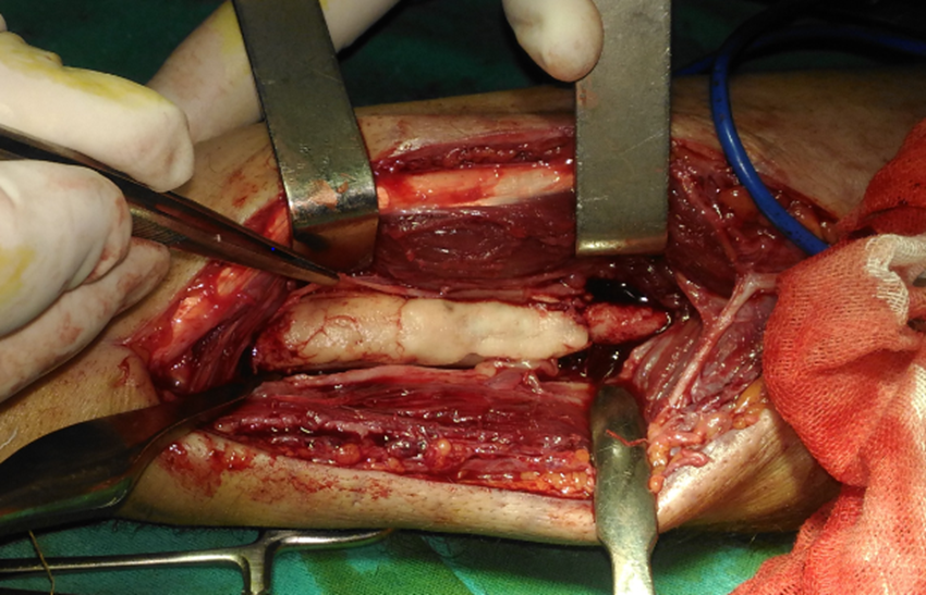
Figure 2. The cement takes the shape of the missing bone segment, and the induced membrane forms around it with the same configuration.
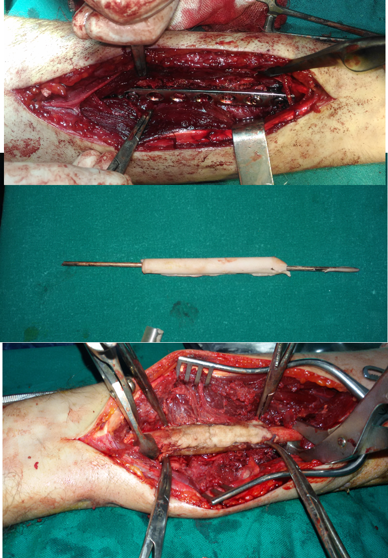
Figure 3. Fixation of the forearm using a compression plate after removal of the bone cement previously stabilised with an intramedullary rod, before proceeding to the second stage and insertion of the definitive bone graft.
Step Five: Adequate Soft-Tissue Coverage
After completing bone fixation and cement placement, another stage of equal importance follows: soft-tissue coverage.
The success of the technique depends to a large extent on adequate coverage of the cement.
Coverage may be achieved by:
-
- Direct wound closure.
- A muscle flap.
- A musculocutaneous flap when there is extensive soft-tissue loss.
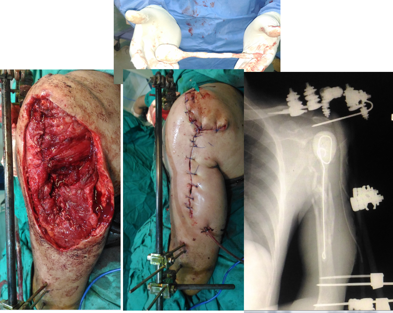
Figure 4. Closure of the wound and surrounding soft tissues in a blast injury of the shoulder, following implantation of cement into the bone defect in the proximal humerus.
What Happens After Completion of the First Stage?
After wound closure, a series of biological changes begins.
During the following weeks:
-
- The induced membrane gradually forms.
- New blood vessel formation increases.
- Growth factor production begins.
- The biological chamber that will subsequently receive the bone graft develops.
Therefore, removal of the cement should not be rushed before the membrane has completed its maturation.
Common Errors During the First Stage
The first stage is extremely sensitive, and certain technical errors may reduce the likelihood of success.
The most important include:
-
- Inadequate debridement, leaving necrotic bone or tissues behind.
- Incomplete cleaning of the infectious focus.
- Unstable bone fixation.
- Application of an insufficient amount of cement or failure to fill the entire defect.
- Inadequate soft-tissue coverage.
- Proceeding to the second stage before confirming that infection is under control.
Avoiding these errors significantly increases the likelihood of successful reconstruction during the second stage.
Conclusion
The first stage of the Masquelet technique is much more than a preliminary procedure. It is the stage in which the biological and mechanical environment required for bone reconstruction is established.
Its success depends on five fundamental principles:
-
- Radical debridement.
- Preparation of the bone ends.
- Stable fixation.
- Correct application of bone cement.
- Adequate soft-tissue coverage.
When these principles are respected, the biological chamber develops over the following weeks, forming the true foundation for successful reconstruction during the second stage.
References
-
- Pelissier P, et al. Induced membranes secrete growth factors including vascular and osteoinductive factors. J Orthop Res. 2004. PMID: 14656662. DOI: 10.1016/S0736-0266(03)00165-7.
- Aho OM, et al. The mechanism of action of induced membranes in bone repair. JBJS Am. 2013. PMID: 23553294. DOI: 10.2106/JBJS.L.00310.
- Masquelet AC. The concept of induced membrane… 2010. PMID: 19931050. DOI: 10.1016/j.ocl.2009.07.011.
- Review 2021. J Orthop Res. Journal of Orthopaedic Research®. H-index: 234. 2020. DOI: 10.1002/jor.24978.
Further Reading: The Masquelet Technique Series
-
- Part I: The Masquelet Technique for the Treatment of Massive Bone Defects.
- Part II: The Induced Membrane – Histological Structure and Biological Properties.
- Part III: Indications for the Masquelet Technique and Selection of Suitable Patients.
- Part V: The Masquelet Technique for the Treatment of Massive Bone Defects.
- Can Massive Bone Defects Be Reconstructed Using a Hybrid Approach?
Dr. Imad Al Hariri
Consultant Orthopaedic Surgeon