
Part III: Indications for the Masquelet Technique and Selection of Suitable Patients
By Dr. Imad Al Hariri
Consultant Orthopaedic Surgeon
Based on a lecture on the importance of the Masquelet technique in the reconstruction of massive bone defects, delivered at the Annual Orthopaedic Surgery Conference, 2018, Omayyad Hotel, Damascus.
Article type: Surgical Technique – Narrative Review
Introduction
The Masquelet technique is no longer limited to the treatment of a single type of bone defect. Over the past three decades, it has become one of the most important bone reconstruction techniques in modern orthopaedic surgery.
It has demonstrated successful outcomes in the treatment of a wide spectrum of complex conditions, ranging from bone defects following open fractures, through nonunions and bone infections, to defects resulting from tumour resection.
The success of this technique depends largely on appropriate patient selection. Understanding the indications, assessing the condition of the soft tissues, controlling infection, and achieving adequate fixation are all factors that directly influence the final outcome.
Contents
-
- How Have the Indications for the Technique Evolved?
- First: Bone Defects Following Open Fractures
- Second: War Injuries and Gunshot Wounds
- Third: Nonunion
- Fourth: Chronic Osteomyelitis
- Fifth: Bone Defects Following Tumour Resection
- Sixth
- How Have the Indications for the Technique Evolved?
How Have the Indications for the Technique Evolved?
When Alain-Charles Masquelet first described this technique, its primary purpose was to treat septic nonunions associated with bone loss following debridement of infected tissues.
Over time, clinical results demonstrated the success of the technique in other, more complex conditions. It is now used in most types of segmental bone defects, whether resulting from trauma, tumours, infections, or congenital deformities.
1. Bone Defects Following Open Fractures
High-energy open fractures are the most common indication for the use of the Masquelet technique.
These injuries are typically associated with:
-
- Direct loss of a segment of bone.
- Severe muscle damage.
- Injury to the skin and soft tissues.
- Varying degrees of bacterial contamination.
In such cases, direct bone grafting is difficult because of the high risk of infection and the absence of a suitable biological environment for graft incorporation.
The Masquelet technique therefore allows the surgeon to:
-
- Perform thorough debridement of the injury site.
- Achieve stable fracture fixation.
- Preserve the bone defect space.
- Prepare a suitable biological environment before performing bone grafting.
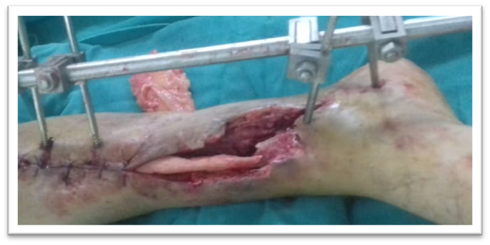
Figure 1. Open fracture with bone loss before treatment.
2. War Injuries and Gunshot Wounds
War-related injuries are characterised by:
-
- Extensive bone loss.
- Severe soft-tissue destruction.
- High levels of contamination.
- A significant risk of infection.
For these reasons, the Masquelet technique has become one of the most frequently used techniques in centres treating war injuries, as it allows treatment to be performed in two stages:
-
- Stage One: Control of contamination and infection, while preserving the bone defect space.
- Stage Two: Reconstruction after improvement of the soft tissues and stabilisation of the patient’s general condition.
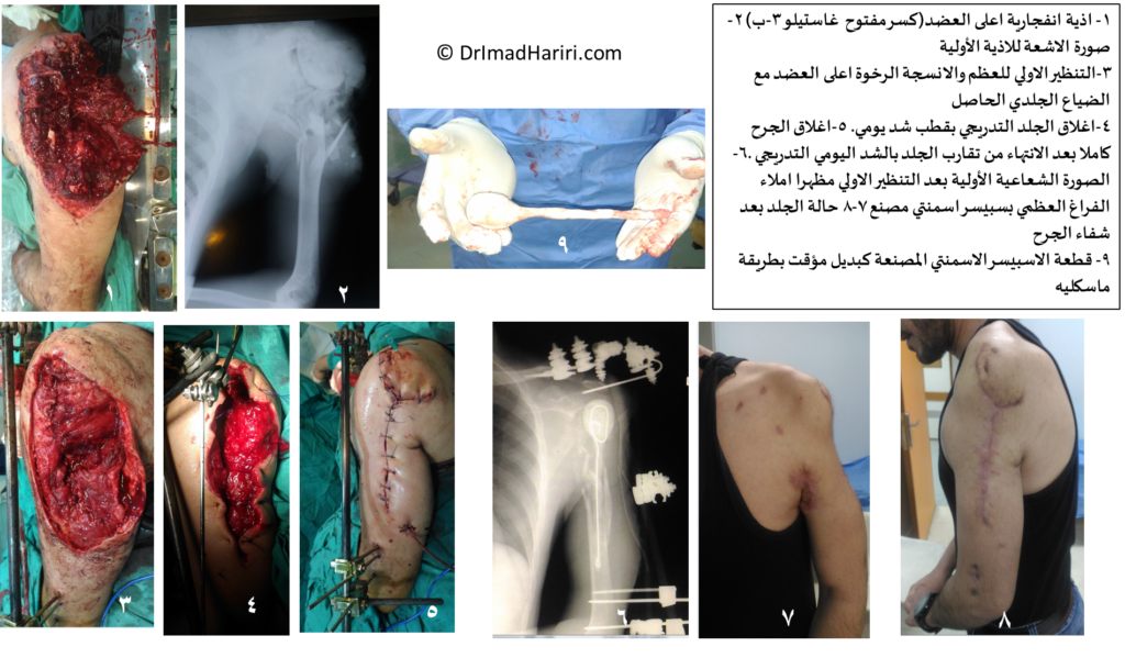
Figure 2. Use of the Masquelet technique in the initial management of open fractures associated with extensive bone and soft-tissue defects.
3. Nonunions
Nonunions, whether septic or aseptic, represent one of the important indications for this technique.
In these cases, treatment is not limited to achieving fracture union. It must also involve:
-
- Removal of fibrous tissue.
- Resection of necrotic bone.
- Reconstruction of the bone defect resulting from debridement.
The induced membrane provides a suitable environment for reconstructing the missing bone segment after the underlying cause of nonunion has been eliminated.
4. Chronic Osteomyelitis
In cases of chronic osteomyelitis, thorough surgical debridement often results in the creation of a substantial bone defect.
This is where one of the major advantages of the Masquelet technique becomes apparent, as it allows the surgeon to:
-
- Remove all necrotic tissues.
- Completely eliminate the infectious focus.
- Insert bone cement, which may be loaded with antibiotics depending on the clinical situation.
- Delay bone grafting until infection control has been confirmed.
The article emphasises that progression to the second stage should not occur until clinical and laboratory evidence indicates that the infection has resolved. Assessment should be based on clinical findings and laboratory tests such as WBC, ESR, and CRP.
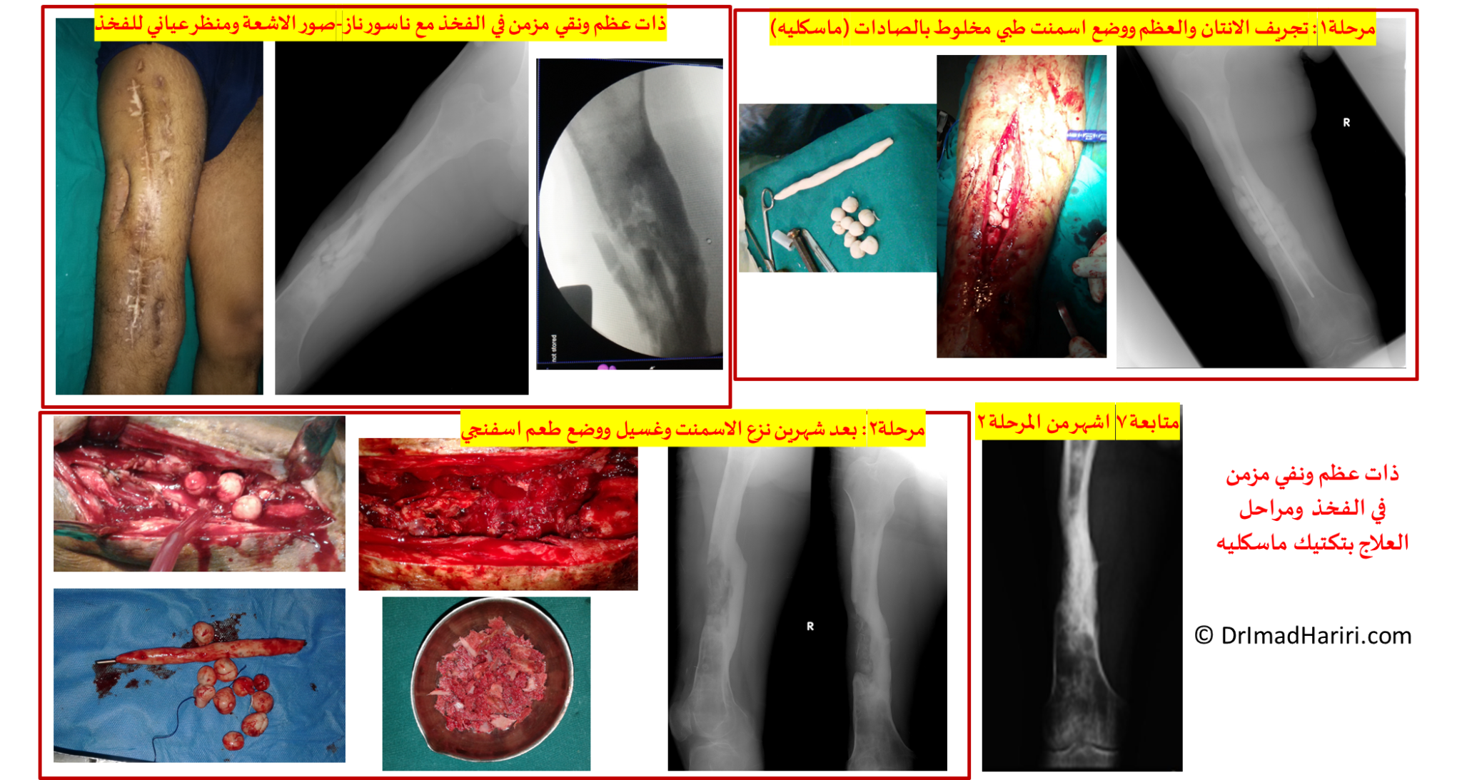
Figure 3. A case of chronic osteomyelitis before and after debridement and application of the Masquelet technique.
5. Bone Defects Following Tumour Resection
The use of the Masquelet technique has become increasingly common in bone tumour surgery, particularly following wide resection of aggressive benign or malignant tumours.
After tumour removal, a large bone defect may remain that cannot be reconstructed using conventional bone grafts.
The technique allows the surgeon to:
-
- Preserve limb length.
- Reconstruct the bone gradually.
- Avoid certain amputation procedures in appropriately selected cases.
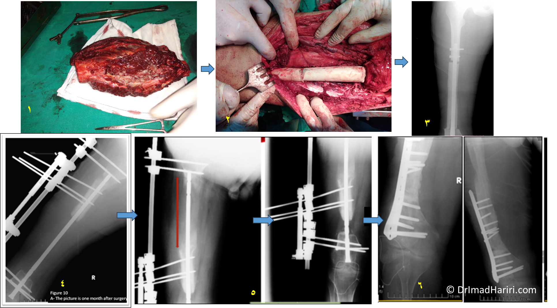
Figure 4. Reconstruction of a bone defect following resection of a malignant bone tumour.
The clinical sequence illustrated in the original figure is as follows:
-
- The resected femoral tumour.
- Placement of cylindrical bone cement within the defect created after tumour resection, with a retrograde femoral nail inserted inside it.
- Radiograph showing the bone cement and the retrograde nail.
- Six months later, removal of the bone cement and insertion of a telescopic intramedullary nail.
- Eight months after the beginning of the lengthening process, showing filling of more than 80% of the bone defect.
- Grafting of the remaining 2.5 cm bone defect using autologous bone graft, with stabilisation using an anatomical distal femoral plate. The radiograph was taken seven months after the final bone grafting procedure.
6. Congenital Pseudarthrosis in Children
One important indication for the Masquelet technique is the treatment of congenital pseudarthrosis in children, particularly following failure of previous treatment attempts.
In these cases, the technique may help reconstruct the bone while maintaining the biological environment required for healing, taking into account the particular characteristics of growing bone and the need for careful treatment planning.
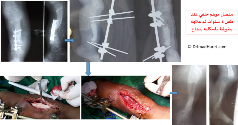
Figure 5. A case of congenital pseudarthrosis of the tibia in a child treated using the Masquelet technique.
Can the Technique Be Used in the Presence of Infection?
One of the concepts that has changed with the development of this technique is that infection is not an absolute contraindication to its use.
On the contrary, the technique can be used following thorough surgical debridement, provided that the fundamental principles of treatment are respected.
These principles include:
-
- Removal of all necrotic tissues.
- Debridement of the bone until viable tissue is reached.
- Stable bone fixation.
- Adequate soft-tissue coverage.
- Delaying bone grafting until the signs of infection have resolved.
This point is emphasised as an essential requirement for the success of the second stage.
Is There a Maximum Length for a Bone Defect?
The Masquelet technique can be used to reconstruct very large bone defects, potentially reaching approximately 30 cm when appropriate surgical conditions are present.
This reflects the considerable reconstructive potential of the technique. Nevertheless, each case must be assessed individually, taking into account:
-
- Blood supply.
- Quality of the surrounding soft tissues.
- Stability of fixation.
- The patient’s general condition.
When Is the Masquelet Technique an Appropriate Option?
The main clinical situations in which this technique may be appropriate are summarised in the following table:
| Clinical condition | Suitability of the technique |
|---|---|
| Bone defects following open fractures | Excellent |
| War injuries and gunshot wounds | Excellent |
| Nonunions associated with bone loss | Excellent |
| Osteomyelitis following debridement | Excellent, after infection control |
| Bone defects following tumour resection | Very good |
| Congenital pseudarthrosis in children | Suitable in selected cases |
Factors Influencing Successful Patient Selection
Before deciding to use the Masquelet technique, several key factors should be assessed, including:
-
-
-
-
-
- The cause of the bone defect.
- The presence or absence of infection.
- The quality of the surrounding soft tissues.
- The possibility of achieving stable bone fixation.
- The patient’s general condition and ability to tolerate staged treatment.
- The availability of a sufficient quantity of bone graft for the second stage.
-
-
-
-
The success of the technique does not depend solely on the size of the bone defect. It also depends on the precise application of all surgical principles and the selection of the appropriate patient at the appropriate time.
Conclusion
The Masquelet technique has demonstrated its ability to treat a wide spectrum of complex bone defects, ranging from open fractures and war injuries to nonunions, bone infections, defects following tumour resection, and certain congenital conditions.
Appropriate patient selection, together with infection control, stable fixation, and preservation of the soft tissues, represents one of the most important factors determining the success of bone reconstruction using this technique.
References
-
- Pelissier P, et al. Induced membranes secrete growth factors including vascular and osteoinductive factors. J Orthop Res. 2004. PMID: 14656662. DOI: 10.1016/S0736-0266(03)00165-7.
- Aho OM, et al. The mechanism of action of induced membranes in bone repair. JBJS Am. 2013. PMID: 23553294. DOI: 10.2106/JBJS.L.00310.
- Masquelet AC. The concept of induced membrane… 2010. PMID: 19931050. DOI: 10.1016/j.ocl.2009.07.011.
- Review 2021. J Orthop Res. Journal of Orthopaedic Research®. H-index: 234. 2020. DOI: 10.1002/jor.24978.
Further Reading: The Masquelet Technique Series
-
- Part I: The Masquelet Technique for the Treatment of Massive Bone Defects.
- Part II: The Induced Membrane – Histological Structure and Biological Properties.
- Part IV: The Masquelet Technique for the Treatment of Massive Bone Defects.
- Part V: The Masquelet Technique for the Treatment of Massive Bone Defects.
- Can Massive Bone Defects Be Reconstructed Using a Hybrid Approach?
Dr. Imad Al Hariri
Consultant Orthopaedic Surgeon